04-06-2024
ADA MED SUPPLY LIMITED
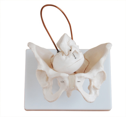
In medical education and clinical practice, the female pelvis and pelvic floor muscle model serves as an intuitive teaching tool that provides learners with an in-depth understanding of the anatomy of the female pelvis. The following is a detailed analysis of the anatomy of this model.

1. Female pelvic structure
The female pelvis is made up of the sacrum, tailbone and two hip bones. The sacrum is located at the lower end of the spine and joins the tailbone and hip bones to form the back of the pelvis. The hip bone includes ilium, ischium and pubis, among which the Angle between the ischium and pubis is particularly important for female childbirth, and its size directly affects the difficulty of childbirth.
On the model, these bone structures are precisely simulated, allowing learners to visually observe their shape, position, and interrelationships. Through touch and observation, learners can gain a deeper understanding of the structural characteristics of the female pelvis.
Second, pelvic floor muscle group
The pelvic floor muscle group is an important group of muscle tissues located at the bottom of the pelvis, which plays a vital role in maintaining the normal position and function of the pelvic organs. The pelvic floor muscle group mainly includes the levator ANI muscle, the coccygeal muscle, the deep transverse perinei muscle and the superficial transverse perinei muscle.
In the female pelvis and pelvic floor muscle model, these muscles are carefully simulated, and their starting point, stopping point and function are clearly demonstrated. For example, the levator anal muscle is located at the bottom of the pelvic cavity, running from both sides of the pelvis and ending in the central tendon of the perineum. Its main function is to support the pelvic organs and assist in defecation. The coccygeal muscle starts from the spine of the ischial bone and ends at the lateral edge of the coccygeal bone. Its main function is to support the internal organs and defecate.
By observing and manipulating the model, learners gain insight into the position, shape, and function of these muscles, as well as their role in female physiological and pathological processes. At the same time, the model can also help learners understand the impact of injury and dysfunction of pelvic floor muscle groups on women's health and how to restore their function through exercise and rehabilitation.
In summary, the female pelvis and pelvic floor muscle model is an intuitive and practical teaching tool that can help learners gain an in-depth understanding of the anatomy of the female pelvis and the functional characteristics of the pelvic floor muscle groups. By learning and mastering this knowledge, learners can better understand the physiological and pathological processes of the female reproductive system and lay a solid foundation for future medical practice.